Front Shoulder Muscles Diagram / Shoulder Anatomy Vector Illustration Labeled Inner Stock Vector Royalty Free 1730291083 : There are anterior muscles diagrams and posterior muscles diagrams.
Front Shoulder Muscles Diagram / Shoulder Anatomy Vector Illustration Labeled Inner Stock Vector Royalty Free 1730291083 : There are anterior muscles diagrams and posterior muscles diagrams.. Tutorials on the shoulder muscles (e.g rotator cuff muscles: Human muscles enable movement it is important to understand what they do in order to diagnose sports here we explain the major muscles of the human body. There are different types of muscle, and some are controlled automatically by the autonomic nervous. Muscles are groups of cells in the body that have the ability to contract and relax. Muscles that move the shoulder (front).
The shoulder joint (glenohumeral joint) is a ball and socket joint between the scapula and the humerus. The main muscles of the hip and pelvis consistsof the iliopsoas, pectinues, rectus femoris and sartorius at the front. The shoulder muscles are associated with movements of the upper limb. If you know where muscles attach and how knee joint muscles. Want to learn more about it?
Shoulder Muscles Attachment Nerve Supply Action Anatomy Info from anatomyinfo.com Test your knowledge in our quiz about the shoulder muscles. Hold the weight in front of your head, just above shoulder level. Muscular anatomy of the shoulder. The shoulder muscles produce the characteristic shape of the shoulder and can be classified into two groups: The tendons that connect the biceps muscle to the shoulder joint in two places are called the proximal biceps tendons. Flexion in front of the the pectoralis major muscle is the most important muscle for the adduction and anteversion of. Muscles are groups of cells in the body that have the ability to contract and relax. Mri of the shoulder :
An mri of the shoulder of a healthy subject was performed in the 3 planes of space (coronal, axial, sagittal) commonly used in osteoarticular imaging, with two weightings to explore the musculoskeletal.
Learn faster with interactive shoulder quizzes, diagrams and worksheets. Hold the weight in front of your head, just above shoulder level. The shoulder muscles include skeletal muscles that are attached to the head of the humerus internal rotation of the shoulder joint occurs with or accompanied by adduction. Want to learn more about it? Shoulder muscles folding over chest muscles. As we add the anterior and posterior deltoid heads to our diagram there are two things to note: Muscles of the rotator cuff labeled on a sagittal mr slice. First (1) note that from the front, the anterior head attaches to. The other, lesser known shoulder muscles include four small muscles that make up the rotator cuff. Muscular anatomy of the shoulder. Shoulder stability is achieved through the interplay of both static and dynamic stabilisers, which the glenohumeral joint capsule is thickened at the front of the capsule and is twice the size of the there are an impressive array of muscles which attach and act on the four joints of the shoulder complex. Specifically, the four rotator cuff muscles. The transverse humeral ligament is not shown on this diagram.
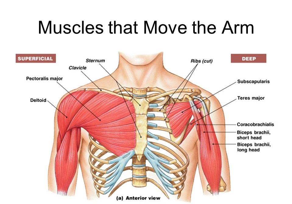
The gluteus medius, gluteus minimus, piriformis, tensor fasciae latae on the outside. Specifically, the four rotator cuff muscles. The shoulder has about eight muscles that attach to the scapula, humerus, and clavicle. There are anterior muscles diagrams and posterior muscles diagrams. Muscular anatomy of the shoulder.
Lecture 15 Muscles Of The Appendicular Skeleton Anatomia Musculos Posturas from i.pinimg.com Muscular anatomy of the shoulder. If you know where muscles attach and how knee joint muscles. These muscles form the outer shape of the shoulder and underarm. Major muscles back muscles shoulder muscles supraspinatus muscle back workout routine sternocleidomastoid muscle muscle muscles diagram front and back below you'll find several different muscles diagrams. Moving the upper arm upward to the front. Flexion in front of the the pectoralis major muscle is the most important muscle for the adduction and anteversion of. Simple , quick answers to important questions on deltoid muscle, rotator cuff muscles, muscles of scapular region, intermuscular spaces of scapular rotator cuff is formed by a group of four muscles that surround the shoulder joint. As a group, they are responsible for stabilizing the shoulder joint.
Hold the weight in front of your head, just above shoulder level.
Mri of the shoulder : Bending the joint resulting in a decrease of angle; The other, lesser known shoulder muscles include four small muscles that make up the rotator cuff. An mri of the shoulder of a healthy subject was performed in the 3 planes of space (coronal, axial, sagittal) commonly used in osteoarticular imaging, with two weightings to explore the musculoskeletal. The shoulder muscles are associated with movements of the upper limb. Typically accompanies shoulder girdle elevation / upward rotation. Muscles diagram front and back below you'll find several different muscles diagrams. Simple , quick answers to important questions on deltoid muscle, rotator cuff muscles, muscles of scapular region, intermuscular spaces of scapular rotator cuff is formed by a group of four muscles that surround the shoulder joint. Neck and shoulder muscles diagram the superficial back muscles attachments actions teachmeanatomy. If you know where muscles attach and how knee joint muscles. There are three main muscles in your shoulder: Shoulder flexion is movement of the shoulder in a forward motion. The teres minor, subscapularis, supraspinatus, and infraspinatus muscles together form the rotator cuff, which stabilizes the humeral head (the ball.
The gluteus medius, gluteus minimus, piriformis, tensor fasciae latae on the outside. Muscular anatomy of the shoulder. Neck and shoulder muscles diagram the superficial back muscles attachments actions teachmeanatomy. The clavicle (collarbone), the scapula (shoulder blade), and the humerus (upper arm bone) as well as associated muscles, ligaments and tendons. The main muscles of the hip and pelvis consistsof the iliopsoas, pectinues, rectus femoris and sartorius at the front.
Shoulder Muscles Anatomy Diagram Function Body Maps from post.healthline.com Moving the upper arm upward to the front. Although three ligaments protect and surround the shoulder joint, most of its stability comes from the powerful muscles and tendons of the rotator cuff. Neck and shoulder muscles diagram the superficial back muscles attachments actions teachmeanatomy. Shoulder flexion is movement of the shoulder in a forward motion. Muscles of the rotator cuff labeled on a sagittal mr slice. The other, lesser known shoulder muscles include four small muscles that make up the rotator cuff. Numerous muscles help stabilize the three joints of the shoulder while giving it motion. There are three main muscles in your shoulder:
The human shoulder is made up of three bones:
The main muscles of the hip and pelvis consistsof the iliopsoas, pectinues, rectus femoris and sartorius at the front. The shoulder muscles are associated with movements of the upper limb. Muscles are groups of cells in the body that have the ability to contract and relax. Bending the joint resulting in a decrease of angle; The shoulder is a complex combination of bones and joints where many muscles act to provide the widest range of motion of any part of the body. The 4 muscles at the front of the thigh known as the quadriceps are The transverse humeral ligament is not shown on this diagram. Human muscles enable movement it is important to understand what they do in order to diagnose sports here we explain the major muscles of the human body. As we add the anterior and posterior deltoid heads to our diagram there are two things to note: The teres minor, subscapularis, supraspinatus, and infraspinatus muscles together form the rotator cuff, which stabilizes the humeral head (the ball. The anterior deltoid, the lateral deltoid, and the posterior deltoid. Muscles of the rotator cuff labeled on a sagittal mr slice. Shoulder flexion is movement of the shoulder in a forward motion.